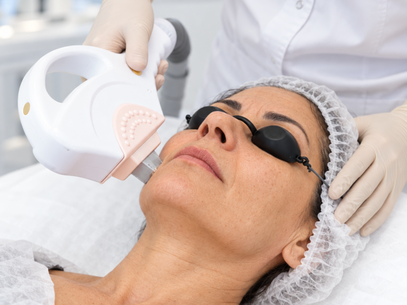


Having many moles is not always an absolute contraindication to IPL, but it does require careful skin examination. Light energy is absorbed by melanin, so a pigmented lesion may be unintentionally heated along with the treatment target. Exposure may also alter the appearance of a nevus and make subsequent monitoring more difficult.
Suspicious lesions must first be assessed by a dermatologist. IPL is not used to diagnose or remove melanocytic lesions and must not obscure signs of possible disease.
What Is Assessed Before Phototherapy?
Before the procedure, the physician examines the entire intended treatment area, not only the vessels or pigment spots. Patients should report any mole that has recently changed shape, color, or size, or has started to bleed.
Dermoscopy allows the structure of a lesion to be examined under magnification. If a lesion is uncertain, phototherapy in that area is postponed until the diagnosis is clarified. When indicated, the physician refers the patient for removal with mandatory histopathological examination.
Which Lesions Require Particular Attention?
Reasons for an unscheduled consultation include asymmetry, irregular borders, uneven color, rapid growth, a new nodule, itching, pain, or bleeding. The “ugly duckling” sign is also important: one mole looks noticeably different from the others.
These signs do not independently confirm melanoma, but they require professional evaluation. A suspicious lesion must not be lightened with IPL, acids, or laser treatment before diagnosis.
How Are Nevi Protected During IPL?
Confirmed benign moles are excluded from the treatment field and covered with an opaque material or carefully avoided with the handpiece. The method depends on their location, size, and the protocol. If lesions are too numerous to treat the area safely, the physician selects another strategy.
Settings also depend on skin phototype and the presence of a tan. Darker skin contains more epidermal melanin, increasing the risk of unwanted heating.
IPL and Skin Cancer Risk
Current reviews have not established that appropriately used cosmetic IPL causes skin cancer. The main clinical concern is exposing an unrecognized suspicious lesion or altering its appearance without histopathological diagnosis.
Safety therefore depends not only on device settings but also on the quality of the pre-procedure examination. The idea that moles can simply be covered is acceptable only after their benign nature has been confirmed.
Monitoring After the Procedure
Pigmented spots may temporarily darken after IPL, but a mole accidentally exposed to a pulse may also change color or develop a crust. This should be reported to the physician promptly, and the crust must not be removed.
Patients with multiple nevi benefit from regular dermatological monitoring and comparison photographs. The specialist determines the appropriate follow-up frequency based on individual risk factors.
How Do Pigment Spots Differ from Moles?
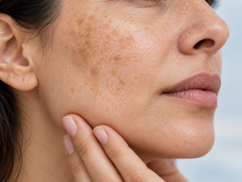
Solar lentigines, freckles, seborrheic keratoses, and melanocytic nevi may all be brown, but they are different lesions. IPL can be used for selected benign pigmentary changes; it must not be applied on the assumption that every dark spot is a treatment target.
Lightening the upper portion of a lesion may make it less visible without removing cells located deeper in the skin. This can create a false impression of treatment and change the appearance used in later diagnostic assessment.
Which Risk Factors Should Be Discussed with the Physician?
A personal or family history of melanoma, numerous atypical nevi, repeated sunburns in the past, and immunosuppression require particular attention. These factors do not always rule out phototherapy completely, but they affect the need for dermatological monitoring.
Patients should report any previous lesion removal and histopathology results. Available records should be brought to the consultation.
What Should You Do If a Mole Is Accidentally Exposed?
The area should not be rubbed, covered with an aggressive adhesive, or treated with acid. Photograph the change and contact the physician. The specialist will assess the reaction and determine the timing of dermoscopic follow-up.
Any crust should separate naturally. After healing, it is important to confirm that the structure and color of the lesion have not changed in a suspicious way.
Preparing the Area for IPL
Moles should not be covered with long-wear makeup or self-tanner before the visit because the physician needs to see the natural skin color. Waxing should be avoided if it irritates the intended treatment area.
When many lesions are present, the consultation may take longer. This should be considered when booking so that a proper examination is not replaced by a brief visual check immediately before treatment.
For a large area, the physician may divide treatment into sections. This allows nevi to be avoided carefully, the skin response to be monitored, and unnecessary increases in energy to be avoided simply to shorten the session.
Before IPL phototherapy at Medest, the physician will examine the area and exclude suspicious lesions. Book a consultation if you have many moles or have noticed a change in any lesion.